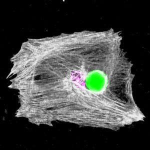
 Our paper on lasers that beat to the rhythm of the heart has just appeared in Nature Photonics (DOI 10.1038/s41566-020-0631-z). In this paper we show how microscopic whispering gallery mode lasers embedded into individual heart cells can be used to report on the contractions of the heart muscle.
60 years after their invention, lasers are widely used in biomedical imaging, resolving ever finer details of life. But because lasers are usually big and power hungry, they tend to sit outside the heart and can only send their light to the surface of the biological tissue, which severely limits how deep they can look. For some time, our group has been interested in embedding micro and nanophotonic devices close to cells of interest to gather more information.
In our latest work, tiny lasers were placed inside the heart where they acted as microscopic probes. With every beat of the heart, the wavelength of the light that these lasers emit changed by a small but clearly detectable amount, thus precisely encoding the contractions of the heart over time.
Marcel Schubert, who is first author on the paper and now runs his own group said, “The wavelength shifts came as a surprise and are believed to be caused by a previously un-recognized change in the cellular machinery of the heart muscle cells.”
We admit that it is still early days, and that more work will be needed until lasers in heart cells can be applied more routinely in other research labs. But the microlasers used can be readily produced in the millions and compared to many modern microscopes, the additional infrastructure required to analyse the laser emission is relatively cheap. To inspire other labs to give this a go, we have made all protocols and the software that analyses the laser output freely available on the university data repository.
